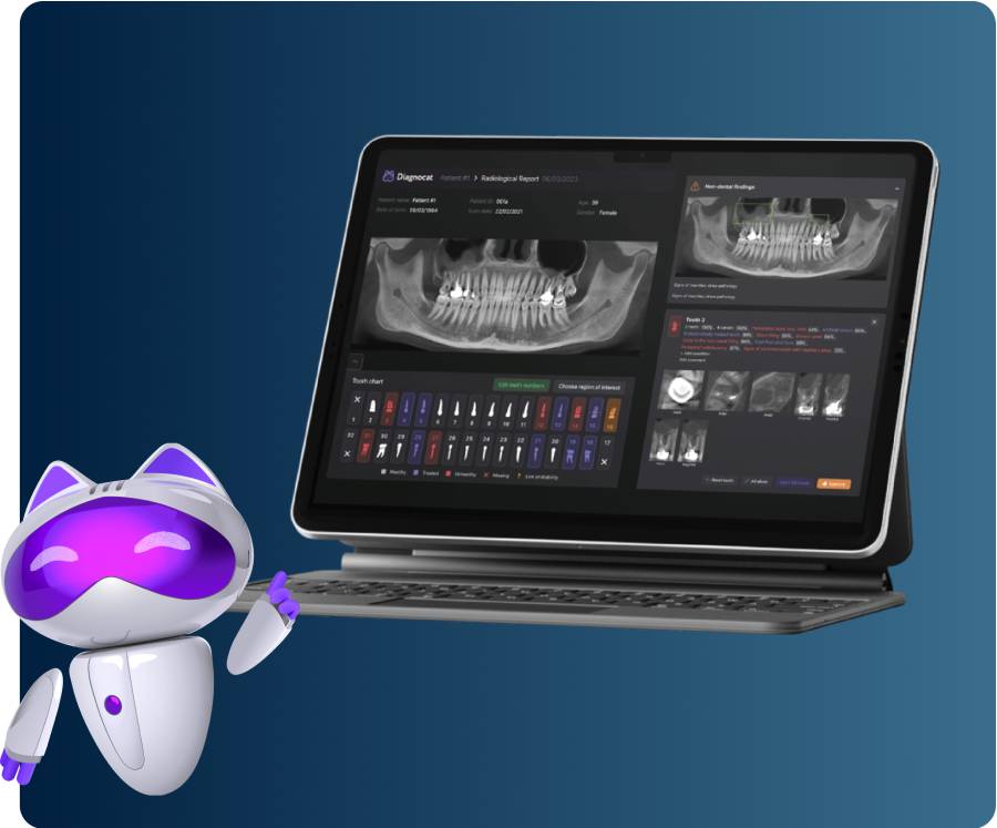
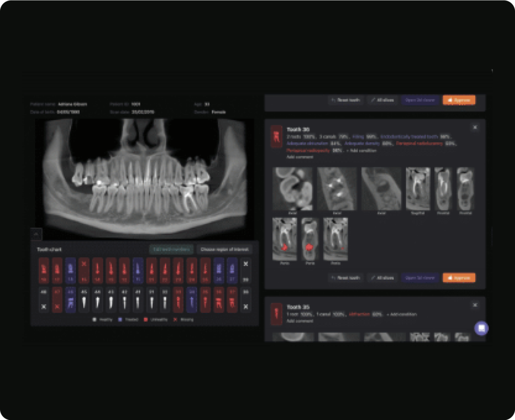
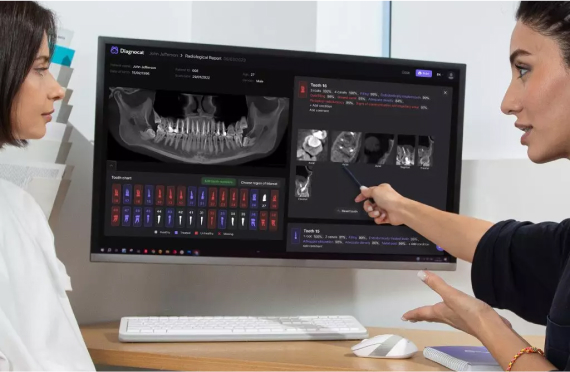
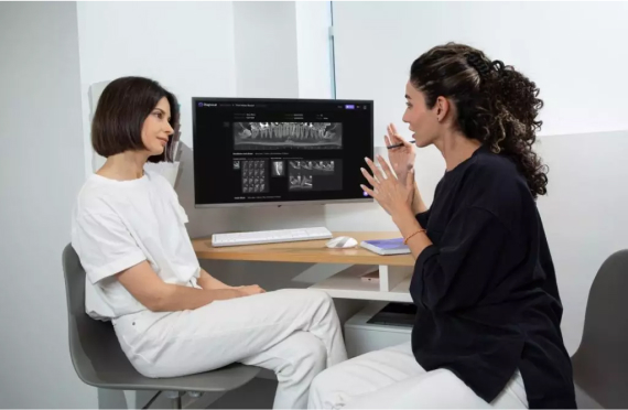
A versatile AI-powered 2D and 3D X-ray analysis system

Discover which solution fits your practice


Radiology Report
Diagnocat’s AI analysis of intraoral X-rays, panoramic X-rays (OPGs), and CBCT images produces an accurate, clear, and concise report of over 65 conditions.
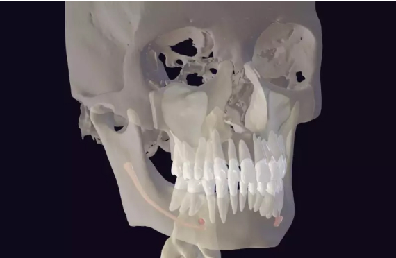
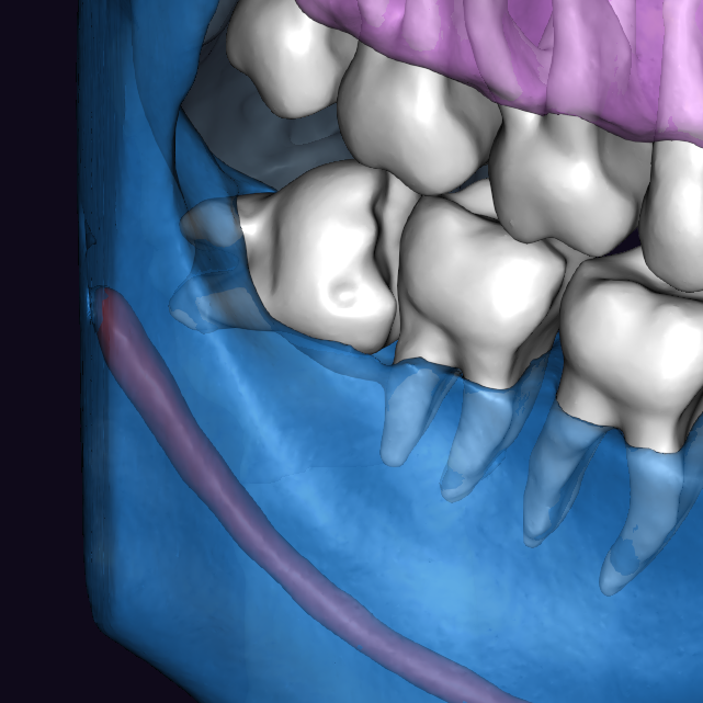
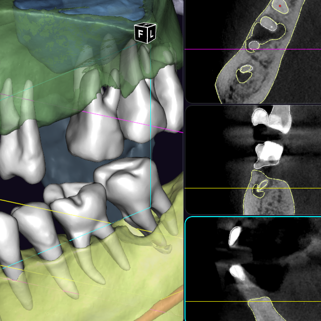
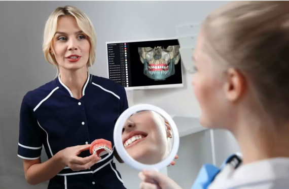
CBCT Segmentation
Diagnocat AI's automatic segmentation feature transforms CBCT files into a 3D STL model, a pivotal innovation for digital dentistry.

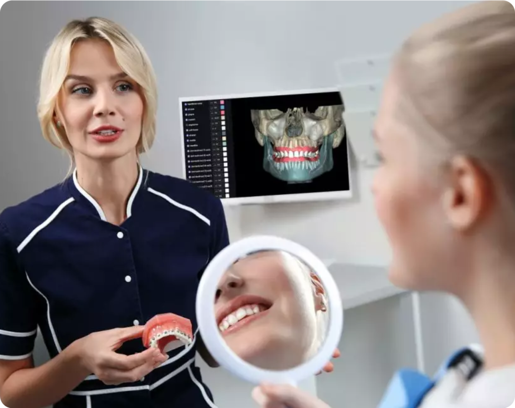
Cloud storage and Viewer
All dental images and reports are securely stored in your cloud-based personal account, accessible for viewing, uploading, sharing, or printing from any device.
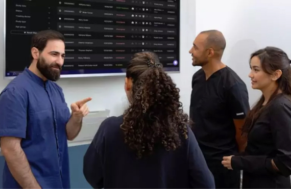
Collaboration Tool
Introducing Diagnocat’s Platform for Comprehensive Treatment Plan Management that transforms our AI into your virtual dental clinic assistant.
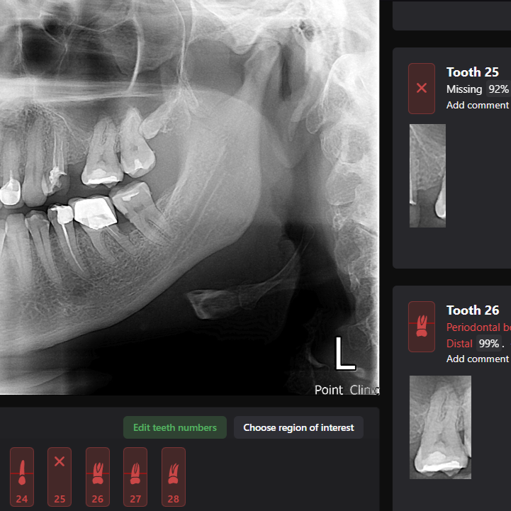
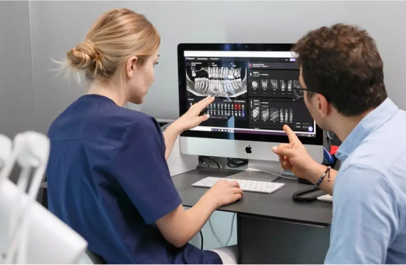
Specialists Reports
Diagnocat offers a range of specialist reports including the third molar, orthodontic and STL reports, easily accessible through the platform.

Superimposition
Our superimposition feature offers dental specialists an enhanced view of their patient's oral cavities by combining the advantages of CBCT and intra-oral imagery.

Diagnocat allows you to create various radiographic reports:
Reports on 2D images are generated based on:
Reports on 3D images are generated based on CBCT.
Additional CBCT-based reports:
Application

Implantology

Wisdom tooth extraction

Orthodontic

Endodontic treatment

STL report

Implantology
Diagnocat uses artificial intelligence to create highly accurate reports for implant planning. The best tools for planning implants are the STL report and the radiological report. The built-in ruler tool allows you to accurately determine the distance to the bottom of the maxillary sinus or to the wall of the mandibular canal.
Key Benefits:
- Detailed cross-sections for bone structure assessment
- Adjustable slice thickness
- Automatic detection of alveolar ridge parameters
- Detailed cross-sections for bone structure assessment
Wisdom tooth extraction
Third molars report by Diagnocat.
Careful planning is the key to reducing the risk of damage to the inferior alveolar nerve during third molar extraction
“Third Molars Report” created by Diagnocat AI is a tool that accurately traces the mandibular canal, builds optimal visualization in three planes and helps the clinician estimate the distance to the mandibular canal
Key Benefits:
- Detailed cross-sections to assess the spatial arrangement of wisdom teeth
- Accurate tracing of the mandibular canal
- Facilitates surgical planning, significantly reducing the risk of complications
- The report is generated in a few minutes
- A detailed and user-friendly report is available for printing or downloading in PDF format


Orthodontic Treatment
Orthodontic report by Diagnocat. The Diagnocat orthodontic report has a fully automatic calculation of 3D cephalometry. Within 5 minutes you receive a ready-made report based on CBCT. 3D cephalometry provides an accurate assessment of anatomical structures, compared to 2D examination, and helps to develop the most effective treatment plan for optimal results.
- The report is generated in a few minutes
- A detailed and user-friendly report, available for printing or downloading in PDF format
The Diagnocat orthodontic report includes
- Panoramic reformat from CBCT
- Summation images from CBCT with outlining of bone structures in AP and lateral projections for the right and left sides
- Cross-sections of the frontal and lateral parts of the teeth
- Coronal sections of the jaws in the area ofmolars and premolars
- Sagittal and coronal sections of the TMJ
- Summation images of sagittal and coronal sections of the TMJ
Endodontic treatment
Key Benefits:
- Detailed sections of the root canal lumen, root canal system, root canal shape, and furcation area.
- Accurate measurements of the periapical lesion volume, which will allow the doctor to evaluate the effectiveness of treatment over time
- Graphic visualization of the periapical lesion helps the patient assess the need for treatment and motivates him to do so.
- Adjustable slice thickness
- The report is generated in a few minutes
- A detailed and user-friendly report is available for printing or downloading in PDF format


STL report
The report generates a 3D CBCT reconstruction, which is convenient for planning orthodontic and surgical treatment, and is also an excellent visualization tool when communicating with the patient
Diagnocat automatically combines intraoral scanning with CBCT, creating a combined model that is actively used in digital dentistry.
The advantages of this report are:
- High quality segmentation of the maxillofacial region
- Built-in multiplanar reconstruction mode with segmentation contours
- The ability to immediately export the combined model for use in digital dentistry
- Fast model creation speed
- Advanced report customization options
Our Webinars
Start using Diagnocat AI
Discover how AI can automate your X-ray analysis and interpretation work.
